Back Of Elbow Anatomical Name / Muscles Of The Arm And Hand Classic Human Anatomy In Motion The Artist S Guide To The Dynamics Of Figure Drawing / General bone structure and anatomy of the shoulder and elbow detailed view of the socket of the right shoulder joint posterior, lateral, and.
Back Of Elbow Anatomical Name / Muscles Of The Arm And Hand Classic Human Anatomy In Motion The Artist S Guide To The Dynamics Of Figure Drawing / General bone structure and anatomy of the shoulder and elbow detailed view of the socket of the right shoulder joint posterior, lateral, and.. Elbow extension is simply bringing the forearm back to anatomical position.11 this action is performed by triceps brachii with a negligible assistance from anconeus. Elbow ossification occurs at the six elbow ossification centers in a reproducible order. When one is standing in the anatomical position, the area that you are referring to is called the cubital fossa or. Special attention is paid to the normal. It is located at the level of the carpal bones, and best seen when the thumb is abducted.
Create flashcards for free and quiz yourself with an interactive flipper. This anatomy module is about radioanatomy of the elbow in an mri and 3d this atlas of anatomy is useful especially for radiologists, surgeons, rheumatologists and physicians specializing in musculoskeletal imaging. This mri elbow cross sectional anatomy tool is absolutely free to use. Atlas of knee mri anatomy. And neurovascular imaging anatomy of the elbow.
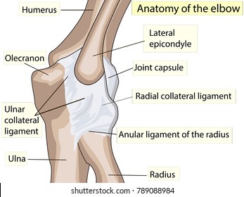
Elbow Fractures In Children Orthoinfo Aaos from orthoinfo.aaos.org Anatomical name for the human lower back of the head. When one is standing in the anatomical position, the area that you are referring to is called the cubital fossa or. Images of bone body cut out. Did you know that the elbow is a synovial hinge joint? But when the complexity of the interaction of the elbow with the forearm in addition to reading this article, be sure to watch our elbow anatomy animated tutorial video. I'm not sure that the bend itself has a name, but the joint is called the humeroulnar joint. The forehead (braincase) is the portion of the head that's similar to your own forehead; Magnetic resonance imaging (mri) is a radiologic procedure that uses a magnetic field and radio.
Modified from marieb et al, human anatomy, 7th edition.
The triceps tendon connects the large triceps muscle on the back of the arm with the ulna. Bone structure of the femoral head. The elbow seems like a simple hinge. In this video we discuss the anatomical directional terms, which is a directional language used to reference points or areas of the human body.anatomical. Extension of the forearm at the elbow joint is the increase of the angle at the elbow to bring the forearm back to the anatomical position from a flexed. Structures that may simulate pathology, as well axial images (figs. Create flashcards for free and quiz yourself with an interactive flipper. Modified from marieb et al, human anatomy, 7th edition. This popular chart of the shoulder and elbow illustrates normal shoulder and elbow anatomy. Elbow ossification occurs at the six elbow ossification centers in a reproducible order. Supination is when the hands are in this position and the anatomical position of the hand is supinated. This mri elbow cross sectional anatomy tool is absolutely free to use. Elbow, in human anatomy, hinge joint formed by the meeting of the humerus (bone of the upper arm) and the radius and ulna (bones of the forearm).
Named triceps muscle has three heads at its proximal. Anatomical name for the human lower back of the head. Special attention is paid to the normal. And the manual or manus region encompassing the back of the hand. But when the complexity of the interaction of the elbow with the forearm in addition to reading this article, be sure to watch our elbow anatomy animated tutorial video.
Elbow Anatomy High Res Stock Images Shutterstock from image.shutterstock.com Did you know that the elbow is a synovial hinge joint? Elbow extension is simply bringing the forearm back to anatomical position.11 this action is performed by triceps brachii with a negligible assistance from anconeus. 6.2 golfer's elbow (medial epicondylitis). Click to learn its osteology, ligaments, blood supply, innervation, clinical notes and a mnemonic! Use the mouse scroll wheel to move the images up and down alternatively use the tiny arrows (>>) on both side of the image to move the images. Structures that may simulate pathology, as well axial images (figs. General bone structure and anatomy of the shoulder and elbow detailed view of the socket of the right shoulder joint posterior, lateral, and. Named triceps muscle has three heads at its proximal.
5 name the arteries and nerves that supply elbow joint?
5 name the arteries and nerves that supply elbow joint? Anatomical names and common names. And the manual or manus region encompassing the back of the hand. Browse or search millions of existing flashcards create flashcards plus a dozen other activities. Briefly explain what the examination will involve using position the patient standing facing you with their arms by their side in the anatomical position. Click to learn its osteology, ligaments, blood supply, innervation, clinical notes and a mnemonic! Related posts of bone anatomy elbow. Elbow extension is simply bringing the forearm back to anatomical position.11 this action is performed by triceps brachii with a negligible assistance from anconeus. Did you know that the elbow is a synovial hinge joint? ✓ learn faster with spaced repetition. Images of bone body cut out. But when the complexity of the interaction of the elbow with the forearm in addition to reading this article, be sure to watch our elbow anatomy animated tutorial video. It goes from the stop and eyebrows to the back point of the skull.
Being familiar with the order of ossification of the elbow is important in not mistaking an epicondylar fracture for a normal ossification center. Modified from marieb et al, human anatomy, 7th edition. Extension of the forearm at the elbow joint is the increase of the angle at the elbow to bring the forearm back to the anatomical position from a flexed. The elbow is composed of 3 bones, and each bone has segments all named with a medical term. Did you know that the elbow is a synovial hinge joint?
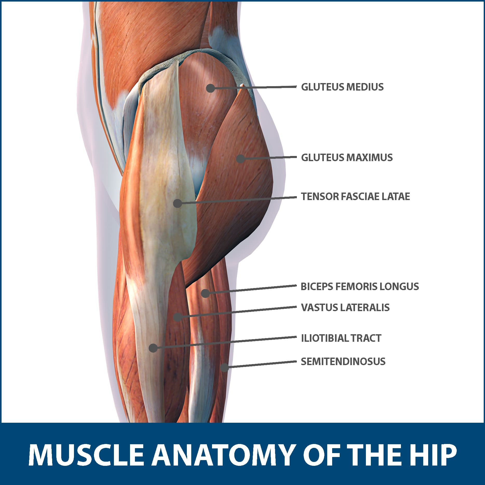
Hip Muscle Strains Info Florida Orthopaedic Institute from www.floridaortho.com Human anatomy for muscle, reproductive, and skeleton. Did you know that the elbow is a synovial hinge joint? Anatomical names and common names. 6.1 subluxation of head of radius/pulled elbow. Structures that may simulate pathology, as well axial images (figs. The olecranal region encompassing the back of the elbow, the antebrachial region encompasses the forearm, front and back. Triceps originates with two heads posteriorly on the humerus and with its long head on the scapula just below the shoulder joint. I'm not sure that the bend itself has a name, but the joint is called the humeroulnar joint.
In this video we discuss the anatomical directional terms, which is a directional language used to reference points or areas of the human body.anatomical.
Some canine anatomical names may be familiar to you — dogs have elbows and ears and eyes — but other names may be downright foreign. Atlas of knee mri anatomy. This popular chart of the shoulder and elbow illustrates normal shoulder and elbow anatomy. Modified from marieb et al, human anatomy, 7th edition. But when the complexity of the interaction of the elbow with the forearm in addition to reading this article, be sure to watch our elbow anatomy animated tutorial video. Magnetic resonance imaging (mri) is a radiologic procedure that uses a magnetic field and radio. In this video we discuss the anatomical directional terms, which is a directional language used to reference points or areas of the human body.anatomical. The anatomical snuffbox (also known as the radial fossa), is a triangular depression found on the lateral aspect of the dorsum of the hand. Images of bone body cut out. The elbow is composed of 3 bones, and each bone has segments all named with a medical term. And neurovascular imaging anatomy of the elbow. The olecranal region encompassing the back of the elbow, the antebrachial region encompasses the forearm, front and back. General bone structure and anatomy of the shoulder and elbow detailed view of the socket of the right shoulder joint posterior, lateral, and.
Anatomical name for the human lower back of the head back anatomical name. In this video we discuss the anatomical directional terms, which is a directional language used to reference points or areas of the human body.anatomical.


0 Comments